Radiology Equipment Management
Modality refers to the specific type of imaging technology used to acquire diagnostic images. In radiology departments the most common modalities include Computed Tomography , Magnetic Resonance Imaging , Ultrasound , Digital Radiography , …
Modality refers to the specific type of imaging technology used to acquire diagnostic images. In radiology departments the most common modalities include Computed Tomography, Magnetic Resonance Imaging, Ultrasound, Digital Radiography, and Nuclear Medicine. Understanding the distinct operational characteristics of each modality is essential for effective equipment management because each device has unique maintenance schedules, calibration requirements, and regulatory obligations. For example, a CT scanner requires routine tube output checks and detector calibration, whereas an MRI system demands strict adherence to magnetic field safety protocols and cryogen management.
DICOM (Digital Imaging and Communications in Medicine) is the universal standard for handling, storing, and transmitting medical images. DICOM defines the file format as well as the network communication protocols that enable interoperability between imaging devices, picture archiving and communication systems (PACS), and radiology information systems (RIS). Proper DICOM configuration ensures that images are correctly tagged with patient identifiers, study information, and acquisition parameters. A common challenge is the occurrence of “DICOM mismatch” errors when a device is upgraded without updating its DICOM settings, leading to lost or misfiled studies and increased turnaround time.
PACS is the central repository that stores and provides access to imaging data across the enterprise. Effective PACS management involves monitoring storage capacity, ensuring network bandwidth, and validating that image retrieval times meet clinical expectations. For instance, a radiology department that experiences frequent PACS downtime may see a decline in diagnostic productivity, as radiologists must wait for images to load before interpretation. Strategies to mitigate these issues include implementing redundant storage arrays, performing regular performance testing, and establishing clear service level agreements (SLA) with the vendor.
Quality Assurance (QA) is a systematic process that verifies imaging equipment is operating within defined performance specifications. QA programs typically incorporate daily, weekly, monthly, and annual tests that assess parameters such as image uniformity, spatial resolution, and contrast-to-noise ratio. In practice, a QA technologist might use a phantom to measure CT number accuracy each month, documenting the results in a logbook. Challenges arise when staffing constraints limit the frequency of QA testing, or when documentation is incomplete, making it difficult to demonstrate compliance during accreditation surveys.
Preventive Maintenance (PM) is scheduled service performed to reduce the likelihood of equipment failure. PM activities include cleaning of components, lubrication of moving parts, and replacement of consumables before they reach end of life. For example, a digital X‑ray system may require quarterly cleaning of the detector panel to prevent dust accumulation that can degrade image quality. The key challenge in PM planning is balancing the need for equipment availability with the downtime required for maintenance, especially in high‑throughput departments where each hour of downtime translates into lost revenue.
Calibration is the process of adjusting equipment to ensure accurate and repeatable performance. Calibration activities are distinct from routine QA checks; they often involve the use of reference standards and may require a service engineer’s intervention. A common calibration task for a PET/CT scanner is the adjustment of the time‑of‑flight correction to maintain quantitative accuracy of tracer uptake values. Improper calibration can lead to diagnostic errors, such as underestimation of lesion size on CT, which underscores the importance of documented calibration records.
Service Contract outlines the terms under which a vendor provides maintenance, repair, and support services for imaging equipment. Contracts typically specify response times, parts coverage, and labor rates. When negotiating a service contract, managers must consider the total cost of ownership (TCO) and the impact of contract terms on equipment uptime. A common pitfall is selecting a contract based solely on the lowest price, which may result in longer response times and higher indirect costs due to extended downtime.
Vendor Management encompasses the processes for selecting, contracting, and overseeing equipment suppliers and service providers. Effective vendor management requires clear communication of performance expectations, regular review of service metrics, and escalation procedures for unresolved issues. For instance, a radiology manager may establish quarterly performance reviews with the CT vendor to assess compliance with the agreed SLA, focusing on metrics such as mean time to repair (MTTR) and mean time between failures (MTBF). Challenges include coordinating multiple vendors for complex multi‑modality departments and ensuring that all parties adhere to the same quality standards.
Asset Lifecycle refers to the stages an imaging device passes through from acquisition to disposal. The lifecycle includes procurement, installation, commissioning, routine operation, maintenance, upgrades, and eventual decommissioning. Managing the asset lifecycle effectively helps optimize capital expenditure, extend useful life, and ensure compliance with regulatory requirements. For example, tracking the age and performance trends of a fluoroscopy system can inform decisions about whether to invest in a refurbishment or replace the unit entirely. One major challenge is maintaining accurate inventory records for large equipment fleets, especially when devices are moved between locations or shared across institutions.
Radiation Safety is a core principle governing the protection of patients, staff, and the public from unnecessary exposure to ionizing radiation. In equipment management, this principle translates into regular checks of shielding integrity, dose monitoring, and adherence to the ALARA (As Low As Reasonably Achievable) concept. Practical application includes installing dose‑monitoring software on CT consoles to track cumulative exposure and automatically alert technologists when preset thresholds are approached. A persistent challenge is balancing the need for diagnostic image quality with the imperative to minimize radiation dose, particularly in high‑volume environments where throughput pressures may encourage higher exposure settings.
Dose Management systems collect and analyze radiation dose data across all modalities, providing insights into trends and opportunities for dose reduction. Implementation of dose management requires integration with DICOM Radiation Dose Structured Reports (RDSR) and the RIS to correlate dose metrics with patient demographics. For instance, a dose management dashboard might reveal that pediatric CT exams consistently exceed recommended dose levels, prompting protocol adjustments and additional staff training. Challenges include ensuring data completeness, protecting patient privacy, and maintaining staff engagement with dose optimization initiatives.
Imaging Protocol defines the specific set of acquisition parameters used for a particular clinical indication. Protocols are tailored to achieve optimal image quality while controlling radiation dose and scan time. Managing protocols involves regular review, standardization across scanners, and documentation of any deviations. A practical example is the creation of a low‑dose head CT protocol for follow‑up of sinus disease, which reduces tube current while preserving sufficient resolution for sinus evaluation. Protocol management challenges include variability in technologist preferences, equipment capabilities, and evolving clinical guidelines.
Worklist Management is the process of organizing and assigning imaging studies to technologists and modalities. An efficient worklist reduces patient wait times and improves workflow predictability. Integration of the RIS with modality worklists enables automatic population of patient demographics and study details, minimizing manual entry errors. For instance, a radiology department might implement a “first‑come, first‑served” rule within the worklist engine, ensuring equitable distribution of cases among technologists. Common challenges include handling emergency studies that must be prioritized, and ensuring that worklist updates propagate promptly across networked devices.
Service Level Agreement (SLA) defines the performance standards that a vendor must meet, typically covering response time, repair time, and parts availability. An SLA may stipulate that critical equipment, such as a linear accelerator, must be back online within four hours of a service call. Establishing realistic SLAs requires analysis of historical downtime data, equipment criticality, and the impact of outages on patient care. A frequent difficulty is negotiating SLAs that are both stringent enough to protect the department’s operational needs and feasible for the vendor to fulfill, especially when staffing constraints affect the vendor’s ability to meet rapid response targets.
Acceptance Testing is performed after the installation of new equipment to verify that the device meets the specifications outlined in the purchase agreement. Acceptance testing includes functional checks, safety verifications, and performance measurements. For example, a new MRI system may undergo a series of tests to confirm magnetic field homogeneity, gradient linearity, and RF coil performance before it is released for clinical use. Challenges in acceptance testing often stem from incomplete documentation, insufficient staffing, or pressure to expedite the go‑live date, which can lead to missed defects that later cause costly repairs.
Commissioning is the comprehensive process of preparing an imaging device for routine clinical operation. Commissioning builds upon acceptance testing and includes detailed calibration, protocol development, staff training, and documentation of performance baseline data. In practice, a CT scanner commissioning team may perform a series of dose measurements, image quality assessments, and workflow simulations to ensure the system integrates smoothly with existing PACS and RIS infrastructure. One of the main challenges is coordinating the many stakeholders—engineers, physicists, IT personnel, and clinical staff—to complete commissioning within a tight timeline while maintaining high standards.
Decommissioning involves safely removing an imaging device from service at the end of its useful life. Decommissioning must address regulatory requirements for disposal of hazardous materials, data sanitization, and environmental considerations. For instance, a decommissioned fluoroscopy system may contain lead shielding and electronic components that require specialized recycling processes. A frequent obstacle is the lack of a clear decommissioning plan, which can result in delays, unexpected costs, or non‑compliance with waste disposal regulations.
Equipment Downtime refers to any period when a device is unavailable for clinical use due to scheduled maintenance, unexpected failure, or other interruptions. Downtime directly impacts departmental productivity, patient throughput, and revenue. Monitoring downtime involves tracking the start and end times of each outage, categorizing the cause, and calculating metrics such as mean time to repair (MTTR). A practical approach to reducing downtime is implementing a predictive maintenance program that uses sensor data to anticipate component wear before failure occurs. However, challenges arise in acquiring reliable sensor data, interpreting trends, and securing sufficient budget for proactive interventions.
Utilization Rate measures the proportion of time a device is actively used for patient imaging relative to its total available time. High utilization indicates efficient use of capital assets, while low utilization may signal over‑capacity or misallocation of resources. Utilization is often expressed as a percentage, calculated by dividing the total scanning hours by the total operational hours in a given period. For example, a CT scanner operating 12 hours per day with an average of 8 scanning hours would have a utilization rate of approximately 67 %. Managing utilization involves scheduling optimization, aligning staffing levels, and possibly redistributing cases among modalities to balance load.
Mean Time Between Failures (MTBF) is a reliability metric that predicts the average interval between consecutive equipment failures. MTBF is calculated by dividing the total operating time by the number of failures observed during that period. A higher MTBF indicates a more reliable device. Radiology managers use MTBF to benchmark equipment performance, negotiate service contracts, and plan preventive maintenance. A common difficulty is obtaining accurate failure data, especially when minor incidents are not formally reported, leading to inflated MTBF values that may mask underlying reliability issues.
Mean Time To Repair (MTTR) quantifies the average duration required to restore a device to operational status after a failure. MTTR is derived by summing the total repair times for all incidents and dividing by the number of incidents. Reducing MTTR is a priority for most departments because faster repairs translate into less lost revenue and improved patient satisfaction. Strategies to lower MTTR include maintaining a well‑stocked spare parts inventory, ensuring that service engineers have immediate access to equipment schematics, and establishing clear escalation pathways. Obstacles can include limited vendor response capabilities, complex repair procedures that require specialized tools, and administrative delays in authorizing repair work.
Risk Assessment is the systematic evaluation of potential hazards associated with equipment operation, maintenance, and decommissioning. In radiology, risk assessments address both safety (radiation exposure, electrical hazards) and operational (downtime, data loss) concerns. A practical risk assessment might involve identifying the probability of a power failure affecting a CT scanner, estimating the impact on patient scheduling, and developing mitigation strategies such as uninterruptible power supplies (UPS) and redundant network paths. Challenges include securing stakeholder buy‑in for mitigation measures and quantifying intangible risks such as reputational damage.
Regulatory Compliance ensures that imaging equipment adheres to laws, standards, and guidelines established by governmental and accrediting bodies. In many jurisdictions, compliance includes adherence to radiation safety standards (e.G., IEC 60601‑2‑33 for CT), equipment labeling requirements, and periodic inspections. Failure to maintain compliance can result in fines, loss of accreditation, or legal liability. Radiology managers must stay current with evolving regulations, conduct internal audits, and maintain documentation that demonstrates compliance. A frequent challenge is the fragmented nature of regulatory requirements, which may differ across federal, state, and professional domains, requiring coordinated effort to satisfy all obligations.
Accreditation Standards such as those set by the American College of Radiology (ACR) or the Joint Commission provide benchmarks for quality and safety in imaging services. Accreditation surveys evaluate aspects such as equipment performance, staff qualifications, and quality improvement programs. Maintaining accreditation often necessitates continuous documentation of QA results, staff training records, and equipment maintenance logs. Practical application includes preparing a “radiology quality manual” that compiles all required evidence for the survey team. Common challenges revolve around the resource intensity of preparing for accreditation cycles and the need to sustain improvements beyond the audit date.
Cost of Ownership (COO) encompasses all expenses associated with acquiring, operating, maintaining, and disposing of imaging equipment. It includes capital purchase price, financing costs, installation, service contracts, consumables, energy consumption, and eventual disposal fees. By analyzing COO, managers can make informed decisions about purchasing new technology versus extending the life of existing assets. For example, a cost‑benefit analysis may reveal that upgrading the detector of an older digital radiography unit is more economical than purchasing a brand‑new system, when factoring in the remaining useful life and expected performance gains. A significant challenge is capturing indirect costs such as lost productivity during downtime, which are often difficult to quantify but can substantially affect the overall financial picture.
Budgeting for radiology equipment requires forecasting capital and operational expenses, aligning them with institutional financial constraints, and justifying investments based on clinical need and return on investment (ROI). Effective budgeting involves multi‑year planning, scenario analysis, and stakeholder engagement. A practical budgeting exercise might involve projecting the replacement cycle for a fleet of CT scanners over a five‑year horizon, estimating the associated service contract costs, and presenting the plan to the hospital finance committee. Challenges include unpredictable technology advancements that may render planned equipment obsolete, and competing priorities from other clinical departments.
Capital Expenditure (CapEx) refers to funds allocated for acquiring long‑term assets such as new imaging systems, major upgrades, or facility renovations. CapEx decisions typically require a formal business case, including clinical justification, cost analysis, and projected impact on patient care. For instance, a proposal to purchase a high‑resolution PET/CT scanner may highlight its ability to improve oncologic staging accuracy, thereby attracting more referrals and increasing revenue. One challenge is that CapEx approvals often involve lengthy review processes, during which market conditions may change, potentially affecting equipment pricing or availability.
Operational Expenditure (OpEx) covers ongoing costs required to keep equipment functional, including service contracts, consumables, utilities, and staff salaries. Managing OpEx involves monitoring expense trends, identifying cost‑saving opportunities, and ensuring that operational budgets align with actual usage. A practical example is negotiating a volume‑based pricing arrangement for contrast media, reducing per‑procedure costs as the department’s imaging volume grows. Challenges include fluctuations in energy costs, inflation of service contract rates, and the need to balance cost containment with maintaining high quality of service.
Vendor Service Call is a request for technical assistance triggered when equipment malfunctions or requires routine maintenance. Effective handling of service calls includes accurate problem description, prioritization based on clinical impact, and tracking of response times. An example workflow might involve a technologist logging a service call for a CT gantry error, assigning it a “high” priority, and the vendor’s service team responding within the agreed SLA window. Common issues include miscommunication of the problem’s severity, delays caused by parts back‑order, and insufficient documentation of the resolution for future reference.
Spare Parts Inventory is the collection of components kept on hand to facilitate rapid repair of equipment failures. Maintaining an optimal spare parts inventory requires balancing the cost of holding inventory against the risk of prolonged downtime due to parts unavailability. For a digital radiography system, critical spare parts might include the detector panel, power supply modules, and cooling fans. A practical approach is to conduct a criticality analysis, identifying parts whose failure would cause the greatest disruption, and ensuring they are stocked with a safety stock level. Challenges include forecasting demand for parts with long lead times, managing expiration dates for consumable components, and avoiding excess inventory that ties up capital.
Warranty Management involves tracking the coverage periods for equipment and components, ensuring that repairs are performed under warranty when applicable, and coordinating with vendors to avoid unnecessary out‑of‑pocket expenses. Effective warranty management can result in significant cost savings, especially for high‑value items like MRI magnets. A typical practice is to maintain a warranty database that alerts managers when a warranty is approaching expiration, prompting decisions about extended warranty purchases or replacement planning. Difficulties arise when warranty terms are ambiguous, coverage limits are unclear, or service technicians inadvertently perform non‑warranty work that voids the agreement.
Performance Metrics are quantitative indicators used to assess the efficiency, effectiveness, and quality of radiology equipment management. Common metrics include equipment uptime, mean time between failures, mean time to repair, utilization rate, dose index trends, and patient throughput. By regularly reviewing these metrics, managers can identify areas for improvement, benchmark against industry standards, and demonstrate value to senior leadership. For example, a dashboard displaying a 95 % equipment uptime over the past quarter can be used to justify continued investment in preventive maintenance programs. The challenge lies in selecting meaningful metrics, ensuring data integrity, and translating results into actionable improvement plans.
Imaging Quality encompasses the visual clarity, contrast, spatial resolution, and artifact level of diagnostic images. Quality is influenced by equipment performance, acquisition parameters, and post‑processing algorithms. Maintaining high imaging quality is essential for accurate diagnosis and patient safety. Practical quality control measures include weekly uniformity checks for digital radiography, monthly noise assessments for MRI, and routine phantom studies for CT. A recurring challenge is the trade‑off between dose reduction and image quality, particularly in pediatric imaging where lower radiation doses may increase image noise, requiring careful protocol optimization.
Detector Aging describes the gradual degradation of detector performance over time, manifesting as increased noise, reduced sensitivity, or persistent artifacts. In modalities such as digital radiography and CT, detector aging can significantly impact image quality and diagnostic confidence. Monitoring detector health involves tracking performance metrics, conducting periodic calibrations, and comparing current measurements against baseline values. For instance, a CT detector panel may show a progressive rise in dark current, indicating the need for panel replacement. A key challenge is determining the optimal replacement point, balancing the cost of a new detector against the clinical impact of diminished image quality.
Software Updates are released by vendors to enhance functionality, address security vulnerabilities, and improve performance of imaging equipment. Applying updates in a timely manner is crucial to maintain compliance with cybersecurity standards and to benefit from new features. A practical update schedule might involve testing a new software patch on a non‑clinical workstation before deploying it to the production environment, thereby minimizing the risk of unintended disruptions. Challenges include coordinating updates across multiple devices, ensuring that changes do not alter acquisition protocols, and managing downtime required for installation.
Cybersecurity in radiology equipment focuses on protecting networked devices, data transmission, and stored images from unauthorized access, malware, and data breaches. Measures include firewalls, encryption, regular patching, and user authentication. A real‑world example is the implementation of network segmentation to isolate PACS from the broader hospital network, reducing the attack surface. The primary challenges involve staying ahead of evolving threats, ensuring staff adherence to security policies, and balancing security controls with usability for clinicians who need rapid access to images.
Data Integrity ensures that imaging data remains accurate, complete, and unaltered throughout its lifecycle. Maintaining data integrity is essential for diagnostic confidence, legal compliance, and research use. Practical steps include implementing checksum verification during image transfer, enforcing access controls, and maintaining audit trails. For example, a radiology department may use DICOM‑based integrity checks to confirm that no bits were corrupted during network transmission from the scanner to the PACS. A common obstacle is the complexity of tracing data corruption events, especially when multiple systems and interfaces are involved.
Backup and Recovery strategies protect imaging data against loss due to hardware failure, natural disaster, or cyber‑attack. A robust backup plan typically involves regular snapshots of the PACS database, off‑site storage of image archives, and periodic recovery drills. An example practice is performing nightly incremental backups combined with weekly full backups, storing copies in a secure cloud repository. Challenges include managing the large volume of imaging data, ensuring that backup windows do not interfere with clinical operations, and testing recovery procedures to confirm they meet required recovery time objectives (RTO).
Radiology Information System (RIS) is the clinical information system that manages patient scheduling, exam ordering, reporting, and results distribution. Effective integration of RIS with imaging modalities and PACS streamlines workflow, reduces manual data entry, and improves patient throughput. For instance, a seamless RIS‑modality interface can automatically populate the patient’s demographics on the CT console, eliminating the need for technologists to re‑enter information. Integration challenges often involve mismatched data fields, version incompatibilities, and the need for custom interface development to accommodate unique departmental workflows.
Imaging Workflow describes the sequence of steps from patient registration to image acquisition, interpretation, and report delivery. Optimizing workflow reduces patient wait times, improves staff efficiency, and enhances overall service quality. Practical workflow improvements may include implementing automated patient routing, using voice‑recognition software for report dictation, and employing real‑time status boards to monitor exam progress. A persistent challenge is balancing the desire for speed with the need for thoroughness, ensuring that shortcuts do not compromise safety or diagnostic accuracy.
Turnaround Time (TAT) measures the interval between exam order completion and final report availability. Shorter TAT is associated with higher patient satisfaction and better clinical outcomes. Monitoring TAT involves tracking timestamps at key workflow stages—order entry, image acquisition, radiologist interpretation, and report signing. For example, a department may set a TAT goal of 30 minutes for urgent X‑ray studies and 2 hours for routine CT exams. Challenges in achieving target TAT include staffing shortages, equipment downtime, and bottlenecks in image transfer or reporting systems.
Throughput refers to the number of examinations performed within a given time frame, often expressed as studies per day or per shift. High throughput is indicative of efficient resource utilization but must be balanced against quality and safety considerations. Practical methods to increase throughput include optimizing scheduling blocks, reducing patient preparation time, and employing parallel processing where multiple scanners operate simultaneously. A common difficulty is that increasing throughput without adequate staffing can lead to burnout, higher error rates, and diminished patient experience.
Radiographer (or Technologist) is the professional responsible for operating imaging equipment, positioning patients, and ensuring image quality. Continuous education and competency assessments are essential for maintaining high standards of practice. For equipment management, technologists play a key role in reporting equipment anomalies, performing routine QA checks, and following maintenance protocols. A challenge is ensuring that technologists have sufficient time to complete QA activities without compromising patient scheduling, especially during peak service periods.
Clinical Engineering is the discipline that provides technical support, maintenance, and engineering expertise for medical equipment. In radiology, clinical engineers collaborate with vendors, oversee preventive maintenance programs, and manage equipment upgrades. Practical involvement includes performing quarterly calibration of CT tubes, coordinating the installation of new software patches, and troubleshooting complex hardware failures. One challenge is the limited number of specialized engineers available for high‑technology modalities such as MRI, requiring careful prioritization of service requests.
Radiation Dose Index is a quantitative measure of the amount of radiation delivered during an imaging exam. Common indices include CTDIvol (Computed Tomography Dose Index volume) and DLP (Dose Length Product) for CT, and DAP (Dose‑Area Product) for fluoroscopy. Tracking dose indices enables departments to benchmark performance, identify outliers, and implement dose reduction strategies. For example, a department may set a target CTDIvol for adult chest CT at 5 mGy, monitoring each scan to ensure compliance. Challenges include variability in patient size, differing protocol settings, and the need to balance dose reduction with adequate image quality.
ALARA Principle (As Low As Reasonably Achievable) guides radiation protection practices by encouraging minimization of exposure while preserving diagnostic efficacy. In equipment management, ALARA is operationalized through protocol optimization, use of automatic exposure control, and regular dose monitoring. A practical illustration is adjusting the kVp and mAs settings on a digital radiography unit for pediatric patients to lower dose while maintaining sufficient image contrast. The main challenge is achieving consensus among clinicians, technologists, and physicists on what constitutes “reasonable” in varying clinical contexts.
Shielding involves the installation of barriers made of lead, concrete, or other attenuating materials to protect staff and the public from stray radiation. Proper shielding design is a prerequisite for safe placement of imaging equipment, especially for high‑dose modalities such as CT and fluoroscopy. For instance, a linear accelerator room may require a 2‑foot lead‑lined wall to contain therapeutic radiation beams. Challenges include ensuring compliance with building codes, managing the cost of installation, and conducting periodic inspections to verify that shielding integrity has not been compromised.
Facility Planning encompasses the design and layout of imaging suites, taking into account space requirements, workflow efficiency, and regulatory constraints. Effective planning improves patient flow, reduces equipment movement, and facilitates future upgrades. A practical example is allocating separate “prep” and “scan” zones for MRI to minimize cross‑contamination and streamline patient handling. Common obstacles involve limited physical space, competing demands from other departments, and the need to accommodate both current technology and anticipated future advancements.
Space Allocation determines the amount of physical area dedicated to each imaging modality, including the scanner footprint, control rooms, and support spaces. Accurate space allocation prevents overcrowding, reduces equipment interference, and supports optimal workflow. For example, a high‑field MRI system may require a minimum of 600 sq ft, including a magnet‑room, control console, and patient changing area. Challenges arise when hospitals undergo renovations, as re‑configuring space can disrupt services and necessitate temporary relocation of equipment.
HVAC Requirements (Heating, Ventilation, and Air Conditioning) are critical for maintaining stable temperature and humidity levels, which affect equipment performance and patient comfort. Certain modalities, such as MRI, have stringent HVAC specifications to prevent magnetic field drift and condensation on sensitive components. A practical measure is installing dedicated climate control units for each scanner suite, with continuous monitoring of temperature and humidity. The challenge is ensuring that HVAC systems are reliable, energy‑efficient, and capable of maintaining specifications despite external weather variations.
Electrical Requirements for imaging equipment include power load, grounding, and backup power provisions. High‑performance devices like CT scanners and linear accelerators draw significant power, often requiring dedicated circuits and surge protection. Proper electrical design prevents voltage fluctuations that could damage equipment or cause image artifacts. For instance, a CT suite may be equipped with a dedicated 400 A three‑phase supply and an uninterruptible power supply (UPS) to sustain operation during brief outages. Common challenges involve coordinating with facility engineers, meeting code compliance, and addressing the high cost of electrical upgrades.
Radiation Protection programs encompass policies, training, and engineering controls designed to safeguard personnel from ionizing radiation. Core components include personal dosimetry, lead aprons, and area monitoring devices. In equipment management, radiation protection also involves regular inspection of shielding, verification of interlock systems, and adherence to exposure limits. A practical scenario is conducting quarterly badge audits to ensure that staff doses remain below regulatory thresholds. Challenges include maintaining staff awareness, updating protection measures as new equipment is installed, and documenting compliance for audits.
Imaging Physics provides the scientific foundation for understanding how imaging modalities generate images, how radiation interacts with matter, and how image quality can be optimized. Radiology managers often rely on medical physicists to perform complex QA tests, assess dose calculations, and advise on protocol adjustments. For example, a physicist may evaluate the effect of changing CT reconstruction algorithms on noise texture and dose efficiency. A frequent challenge is ensuring that physics expertise is available on‑site, especially in smaller facilities where the physicist may be shared across multiple departments.
Image Reconstruction is the computational process that converts raw data from scanners into interpretable images. Advances in reconstruction algorithms, such as iterative reconstruction for CT, can improve image quality while reducing radiation dose. Managing reconstruction software involves staying current with vendor releases, validating new algorithms against established standards, and training staff on any workflow changes. A practical illustration is a radiology department adopting model‑based iterative reconstruction, resulting in a 30 % dose reduction for abdominal CT scans. Challenges include the need for additional processing time, compatibility with existing PACS, and ensuring that radiologists are comfortable interpreting images produced by newer algorithms.
Flat Panel Detector technology is used in digital radiography, fluoroscopy, and some CT systems to capture X‑ray images with high spatial resolution and fast acquisition times. Proper management of flat panel detectors includes routine cleaning, protective handling, and timely replacement of aging panels. For example, a fluoroscopy suite may schedule monthly inspections of the detector surface to detect scratches or dead pixels that could degrade image quality. The primary challenge is the high cost of detector replacement, which necessitates careful monitoring of performance trends to predict the optimal replacement timing.
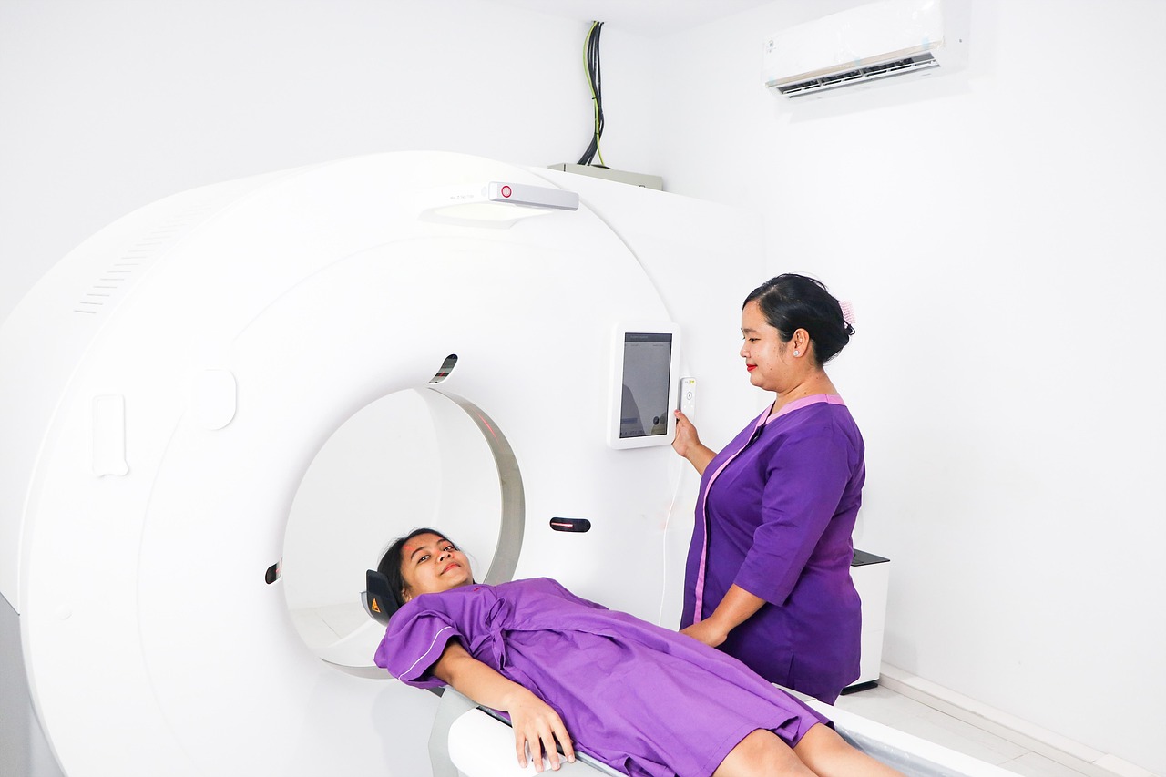
CT Scanner is a high‑speed, high‑resolution imaging modality that acquires volumetric data using rotating X‑ray tubes and detectors. Managing CT equipment involves overseeing tube lifespan, monitoring dose indices, and ensuring that software updates maintain compatibility with PACS. A practical maintenance task is the periodic replacement of the X‑ray tube after a predetermined number of mA‑hours, preventing unexpected failure that could halt scanning operations. Common challenges include managing the high cost of tube replacement, coordinating downtime for service, and maintaining consistent image quality across multiple CT units.
MRI (Magnetic Resonance Imaging) utilizes strong magnetic fields, radiofrequency pulses, and gradient coils to generate detailed soft‑tissue images. MRI equipment management requires attention to magnet cooling systems, RF coil maintenance, and strict safety protocols for ferromagnetic objects. A typical safety measure is conducting a daily “magnet safety check” that verifies the status of the quench system and ensures that the door interlocks are functional. Challenges include the complexity of cryogen handling, the need for specialized training for staff, and the high cost of repairs when gradient coils malfunction.
Ultrasound employs high‑frequency sound waves to create real‑time images, often used for obstetric, vascular, and point‑of‑care applications. Equipment management for ultrasound includes routine probe cleaning, firmware updates, and calibration of depth and gain settings. For instance, a department may implement a weekly test using a standard phantom to verify that the axial resolution remains within acceptable limits. A frequent challenge is the rapid turnover of handheld devices, which can lead to inconsistent quality control practices across the fleet.
Nuclear Medicine involves the administration of radiopharmaceuticals and the acquisition of images using gamma cameras or PET scanners. Managing nuclear medicine equipment requires strict adherence to radiation safety regulations, proper handling of isotopes, and regular calibration of detector energy windows. A practical task is performing a daily uniformity check on a gamma camera using a flood source to detect any degradation in detector response. Challenges include managing the short half‑life of radiotracers, ensuring compliance with stringent licensing requirements, and coordinating with the pharmacy for timely radiopharmaceutical delivery.
PET/CT combines positron emission tomography with computed tomography to provide both metabolic and anatomic information. Equipment management for PET/CT includes monitoring the performance of both the PET detector and the CT subsystem, as well as maintaining the cyclotron or radiopharmacy supply chain for tracer production. A typical QA activity is measuring the PET system’s spatial resolution using a point source phantom, ensuring that the system meets the manufacturer’s specifications. Challenges arise from the complexity of integrating two distinct modalities, the high cost of maintenance, and the need for specialized technical expertise.
Linear Accelerator (Linac) is a device used in external beam radiotherapy to accelerate electrons and generate high‑energy photon beams for cancer treatment. Managing a linac involves regular checks of beam output, mechanical alignment, and safety interlocks. A practical example is performing a monthly output constancy test using a calibrated ion chamber to verify that the radiation dose delivered matches the planned dose. The primary challenges include the need for highly specialized service personnel, strict regulatory compliance, and the impact of any downtime on patient treatment schedules.
Radiotherapy Equipment also includes brachytherapy units, treatment planning computers, and patient positioning systems. Effective management requires coordination between physics, dosimetry, and clinical teams to ensure accurate dose delivery. For instance, a brachytherapy afterloader must be calibrated annually using a well‑characterized source to verify dwell times. Challenges include maintaining a secure inventory of radioactive sources, ensuring that software updates do not alter treatment parameters inadvertently, and managing the high cost of equipment upgrades.
Contrast Injector (or Power Injector) automates the delivery of contrast media during CT, MRI, and angiographic procedures, improving consistency and timing. Management of injectors includes routine calibration of injection pressure, verification of tubing integrity, and adherence to manufacturer‑specified cleaning protocols. A practical maintenance task is the quarterly inspection of the injector’s syringe pump to detect wear that could affect flow rate accuracy. Challenges involve maintaining a stock of disposable tubing sets, ensuring compatibility with a variety of contrast agents, and training staff to recognize injector alarms promptly.
Patient Positioning devices such as positioning aids, laser alignment systems, and motorized tables are essential for reproducible image acquisition. Proper management includes regular inspection for wear, calibration of table motion, and replacement of worn positioning cushions. For example, a CT scanner’s motorized table may require annual verification of travel accuracy to prevent misregistration of images.
Key takeaways
- Understanding the distinct operational characteristics of each modality is essential for effective equipment management because each device has unique maintenance schedules, calibration requirements, and regulatory obligations.
- DICOM defines the file format as well as the network communication protocols that enable interoperability between imaging devices, picture archiving and communication systems (PACS), and radiology information systems (RIS).
- Strategies to mitigate these issues include implementing redundant storage arrays, performing regular performance testing, and establishing clear service level agreements (SLA) with the vendor.
- Challenges arise when staffing constraints limit the frequency of QA testing, or when documentation is incomplete, making it difficult to demonstrate compliance during accreditation surveys.
- The key challenge in PM planning is balancing the need for equipment availability with the downtime required for maintenance, especially in high‑throughput departments where each hour of downtime translates into lost revenue.
- Improper calibration can lead to diagnostic errors, such as underestimation of lesion size on CT, which underscores the importance of documented calibration records.
- A common pitfall is selecting a contract based solely on the lowest price, which may result in longer response times and higher indirect costs due to extended downtime.